
MIKAIA® supports various I/O formats, find out which ones. Importable whole-slide-image file formats Format / VendorCommentsOME-TIFFBrightfield and fluorescence. Z-stacks and timelines supported (mutually-exclusive). No...

![]()
MIKAIA is the ideal software for preclinical and clinical research – precisely tailored to the requirements and specific questions of life science researchers at every level.
MIKAIA® supports various I/O formats, find out which ones. Importable whole-slide-image file formats Format / VendorCommentsOME-TIFFBrightfield and fluorescence. Z-stacks and timelines supported (mutually-exclusive). No...

This MIKAIA® App Note shows how to quantify tumor in proximity to nerve fibers (perineural invasion) in a IHC duplex-stained tissue section, where nerves appear red (antigen: S100) and tumor brown (antigen:...
Digital Pathology is a new data source to transform medicine Medscape – Oct 15, 2014. This was the hook line of a 2014 article in Medscape we stumbled across the other day. Ten years later...
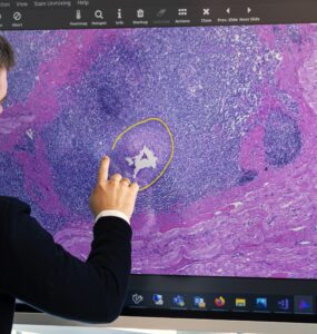
The first step to creating a supervised Digital Pathology AI is to annotate whole-slide-images. MIKAIA® uses various annotation concepts, and understanding these concepts will make your experience with MIKAIA® much...
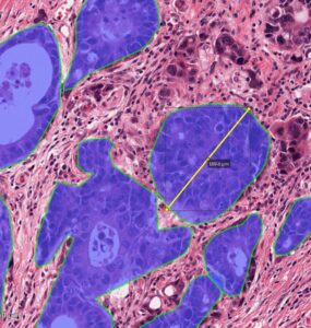
About 20 % to 25 % of breast cancers depend on amplification and overexpression of the gene human epidermal growth factor receptor 2 (HER2). The HER2 overexpression status will initially be assessed by...
The TherVacB project, led by Helmholtz Zentrum München, received a five-year funding within the Horizon 2020 program. With TherVacB, a therapeutic vaccine to cure Hepatitis B, the project’s aim is to provide a...
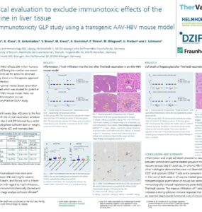
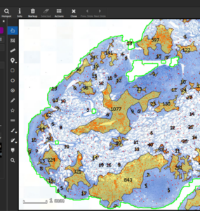
When designing a new immunohistochemistry (IHC) assay, a quantitative assessment of how it “responds” in different tissues of various organs is insightful and helps optimizing the assay in an objective way...
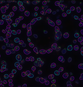
Tumor cells dynamically interact with their microenvironment to stimulate cancer development, progression, and metastasis. To understand the biology of the tumor-immune interactions, it is essential to identify tumoral...
Digital pathology and artificial intelligence (AI) go hand in hand to revolutionize medical diagnostics. However, pathology AI models must be able to cope with a very large variety of histological images if they are to...
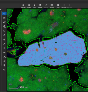
In this MIKAIA® University application note, we guide you through the analysis of a common use case, coexpression of marker combinations in islets of Langerhans and surrounding tissue in a pancreas 4plex imaged with...

© Fraunhofer IIS · 2026