the central MIKAIA® learning portal
get inspired
(note: in February 2024 MICAIA was renamed to MIKAIA)
MIKAIA® University is the central MIKAIA learning portal. Here you can find various App Notes and Video Tutorials that describe typical MIKAIA® workflows either from a technical or medical perspective. As we are continuously adding more content, please check back frequently or subscribe to the MIKAIA newsletter, where we announce newly released App Notes.
MIKAIA University App Notes
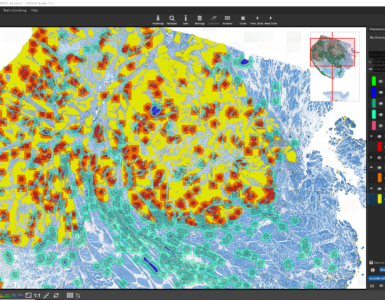
MIKAIA® University App Notes are blog articles, where we describe a particular MIKAIA® app or a medical use case in a step-by-step fashion. Our app notes typically describe what the expected input images look like and what quantitative outputs can be generated. They contain many screenshots and explain the concepts or in some cases even the technical background of the image analysis apps.
We are frequently releasing new app notes.
User Manual
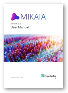
A detailed 90+ pages user manual is included in the free MIKAIA® installer (download here) and available from within MIKAIA® by clicking “User Manual” in the main “Help” menu.
MIKAIA Configurations
Don’t worry about selecting which apps you want to purchase. Currently, three feature sets are available:
- MIKAIA® lite (free)
- MIKAIA® studio
- MIKAIA® studio + AI Author App
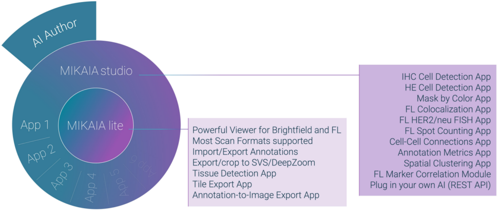
If you’d like to try out the MIKAIA® apps on a temporary basis or request a quote, please feel free to email us at mikaia@iis.fraunhofer.de.
Licensing Options
By default, we offer perpetual licenses, i.e., once activated, features are available forever, without any follow-up costs.
A license can be node-locked, which means it is locked to a particular PC, or alternatively, we offer single-user or multi-user floating licenses. Here, the activation file is imported on a server in your network. MIKAIA can then be installed on any computer in your network or on a (VPN-connected) home office laptop. At startup, MIKAIA® will automatically detect the license server and, if a user-slot of the single- or multi-user license is currently free, it will start up as MIKAIA® studio, else as MIKAIA® lite. The licensing modes are also illustrated in the MIKAIA product brochure.
Working collaboratively
Even though MIKAIA® is not a web-based software, it still can be used collaboratively: The free MIKAIA® lite edition can be used to create annotations manually, but it can also view annotations generated by an analysis conducted with MIKAIA® studio. In other words, user A can run an analysis with MIKAIA studio on computer A, and later users B-Z can view the results on their own computers using the free MIKAIA® lite edition. MIKAIA stores annotations decentrally in a small *.ano file next to the whole-slide-image file:


This is handy, because it enables users to move the slide (incl. *.ano file) into another folder and the connection between slide and annotation will not get broken. Users can also keep multiple versions of annotations, simply by creating copies of the *.ano file, or create backups of annotation files, for instance after they have created manual annotations.
It is also possible to create shortcuts to the slides and then put an *.ano file next to the shortcut. When opening the shortcut, MIKAIA® will open the annotations located next to the shortcut instead of the annotation file located next to the original slide. This way, multiple annotation sets can be maintained, without having to duplicate the (large) WSIs. MIKAIA® has built-in dialogs that can be used to batch-create shortcuts or retarget existing shortcuts (in case the original WSIs have been relocated).
Required Hardware and OS
Currently, MIKAIA® is available for Microsoft Windows, not for Linux or Mac OS.
We offer both an installer and a portable version. The installer is an *.exe file and it requires admin rights for installation. The portable version is a ZIP file that must be manually extracted and then MIKAIA.exe can be started from there. This process does not require admin rights.
AI-enabled image analysis Apps run faster, when a NVIDIA CUDA-enabled GPU is available. We recommend a GeForce card with tensor cores, e.g. GeForce RTX 40xx. When no such GPU is available, the Apps will still work and use the CPU instead.
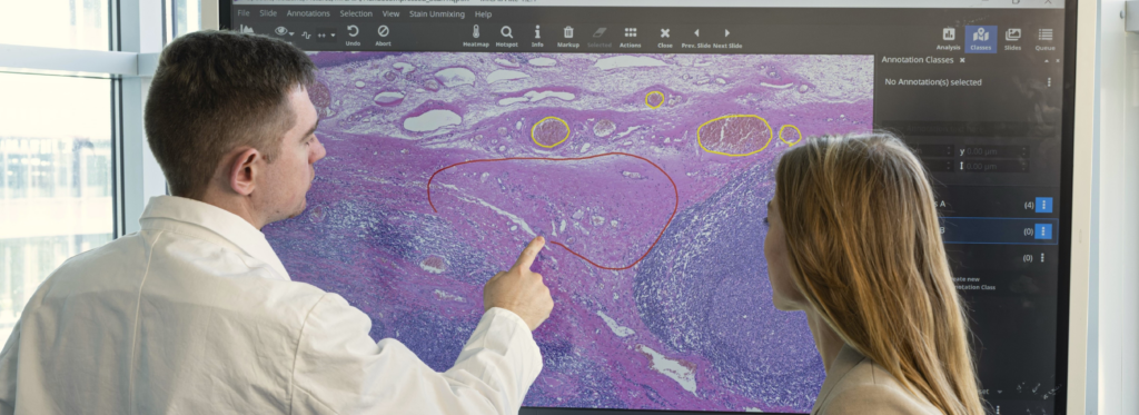
Both 2K and 4K displays are supported. MIKAIA® has also good touch-support. For creating accurate annotations, we recommend using a tablet with a stylus pen (we use WACOM tablets). At exhibitions, we sometimes show MIKAIA® on a touch-enabled digital whiteboard (we use a Samsung Flip 2).
Due to the large size of whole-slide-images, it is recommended to have 16 GB RAM or more.
MIKAIA® University Video Tutorials
These and further videos are available in the MIKAIA® YouTube playlist.





Add comment