
This MIKAIA® University app note demonstrates how to quickly and interactively train your own AI using just a few training annotations, all in a matter of minutes. In the app center, select the Segmentation AI Author...

MIKAIA University Use Case
This MIKAIA® University app note demonstrates how to quickly and interactively train your own AI using just a few training annotations, all in a matter of minutes. In the app center, select the Segmentation AI Author...

A strong suit of MIKAIA® is its wide portfolio of analysis apps for multiplexed immunofluorescence (mIF) / spatial proteomics. It can be used for both low-plex and high-plex panels and is compatbile with image formats...

This MIKAIA® app note demonstrates how to analyze a 47-plex spatial proteomics scan created with MACSima ™ (by Miltenyi Biotec), utilizing sequential immunofluorescence for the analysis: Instrument: MACSima™ by Miltenyi...

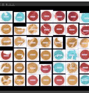
Researchers at the Fraunhofer Institute for Toxicology and Experimental Medicine (ITEM) have developed PEDRA: Platform for Ex-Vivo Drug Response Assays. Up to 100 compounds are tested in parallel on primary patient...

The Laboratory for Bio-Micro Devices at Brigham and Women’s Hospital () develops drug releasing implantable intratumoral microdevices (IMD) [1]. Using MIKAIA®, they want to examine in a quantitative fashion how...

CODEX, now called PhenoCycler, by Akoya Biosciences, along with other sequential immunofluorescence technologies (e.g., Lunaphore (Comet), Miltenyi Biotech (MACSima), Leica Microsystems (Cell DIVE), or other vendors)...

Result of Grid Analysis App shows histological growth pattern and spatial heterogeneity

This article is based on a our presentation “IHC Cell Analysis — More than just Cell Counting: A proposed Workflow” at the European Conference for Digital Pathology (ECDP) 2024 in Vilnius, Lithuania...

This MIKAIA® App Note shows how to quantify tumor in proximity to nerve fibers (perineural invasion) in a IHC duplex-stained tissue section, where nerves appear red (antigen: S100) and tumor brown (antigen:...
When designing a new immunohistochemistry (IHC) assay, a quantitative assessment of how it “responds” in different tissues of various organs is insightful and helps optimizing the assay in an objective way...

© Fraunhofer IIS · 2026