
Chromogenic in situ hybridization (CISH) is used to detect specific DNA or RNA sequences in tissue samples. It uses enzyme-based staining rather than fluorescence (FISH), allowing for high-resolution imaging...

Chromogenic in situ hybridization (CISH) is used to detect specific DNA or RNA sequences in tissue samples. It uses enzyme-based staining rather than fluorescence (FISH), allowing for high-resolution imaging...

This MIKAIA® University app note illustrates how to interactively train a new H&E segmentation AI with serial sections, without having to annotate tumor regions. Instead, an IHC epithelial marker (pan-cytokeratin...

The “Divide by ROIs” feature in MIKAIA’s analysis apps enables users to compile statistics and enhance their understanding of variations in cell populations. This functionality allows for comparisons between user...

The Universal IHC Cell AI App features a universal subcellular IHC cell AI that is compatible with a wide range of markers, tissues, and cell types. Our 23-minute video tutorial guides MIKAIA® step by step through the...

This gallery illustrates for a range of different tissue types and IHC markers the cell analysis quality of MIKAIA‘s IHC Cell Detection App. The app first unmixes the stain components, then uses an AI to delineate...

Result of Grid Analysis App shows histological growth pattern and spatial heterogeneity

Cancer is a major cause of mortality worldwide, and the evaluation of predictive and prognostic markers is crucial in its pathologic assessment. Immunohistochemistry (IHC) is commonly used to evaluate biomarker status...

This article is based on a our presentation “IHC Cell Analysis — More than just Cell Counting: A proposed Workflow” at the European Conference for Digital Pathology (ECDP) 2024 in Vilnius, Lithuania...

This MIKAIA® App Note shows how to quantify tumor in proximity to nerve fibers (perineural invasion) in a IHC duplex-stained tissue section, where nerves appear red (antigen: S100) and tumor brown (antigen:...
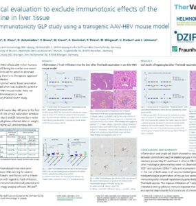
The TherVacB project, led by Helmholtz Zentrum München, received a five-year funding within the Horizon 2020 program. With TherVacB, a therapeutic vaccine to cure Hepatitis B, the project’s aim is to provide a...

© Fraunhofer IIS · 2026