
 Volker Bruns
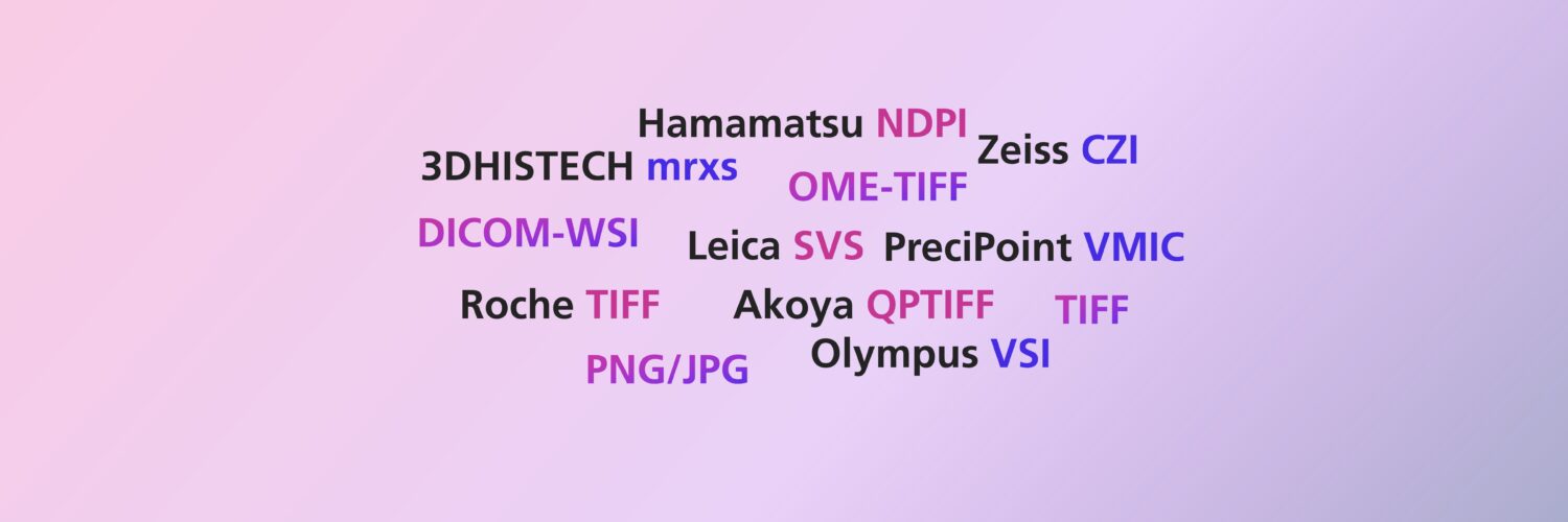
MIKAIA® supports various I/O formats, find out which ones.
Importable whole-slide-image file formats
| Format / Vendor | Comments |
|---|---|
| OME-TIFF | Brightfield and fluorescence. Z-stacks and timelines supported (mutually-exclusive). No multi-file support. |
| DICOM-WSI | Brightfield only |
| Aperio SVS | Brightfield only |
| Aperio SCN | Brightfield & Fluorescence |
| Hamamatsu NDPI & NDPIS | Brightfield and fluorescence |
| 3DHISTECH Mirax | Brightfield and fluorescence (3 channels only) |
| Roche Ventana TIFF | Brightfield only |
| MetaMorph TIFF | Fluorescence only. Z-stacks supported |
| Leica SCN | Brightfield and fluorescence |
| PreciPoint VMIC | Brightfield only |
| Olympus VSI | Brightfield and fluorescence |
| Zeiss CZI | Brightfield and fluorescence. Z-stacks supported Support must be unlocked. Get in touch for further details. |
| Kfbio | Brightfield only. Use *.svs format |
| Perkin Elmer QPI | Brightfield and fluorescence |
| NanoString GeoMX TIFF | Image only (not RNAs) |
| Quanterix / Akoya Biosciences / CODEX QPTIFF (PhenoImager & PhenoCycler) | Fluorescence |
| Lunaphore COMET Ome-TIFF | Fluorescence |
| Milteny MACSima TIFFs | Fluorescence highplex. Channels are single-tiff. They can be loaded as a multiplex, by adding a *.multifile.csv file (refer to MIKAIA user manual) see also: Analyzing MACSima 47-plex mIF with MIKAIA: AI Cell Segmentation + Cell Typing + Cell-cell Connections + Cellular Neighborhoods |
| 10x Genomics Xenium | multi-omics stack comprising “morphology” channel and multiple protein channels. Cell polygons and RNA spots are natively displayed as annotations. Genes are mapped to annotation classes. (since MIKAIA v3.0) see also: Xenium Analysis with the MIKAIA Cell x Gene App |
| Singular Genomics G4x | multi-omics stack comprising H&E channel and multiple protein channels. Cell polygons and RNA spots are natively displayed as annotations. Genes are mapped to annotation classes. (upcoming in MIKAIA v3.1) |
| Keyence TIFF | Brightfield and fluorescence |
| Motic TIFF | Brightfield only |
| ImageJ TIFF | Brightfield only |
| multipe TIFFs (one per channel) | multiple TIFFs (one per channel) can be combined into a multiplex fluorescence scan by placing a CSV file next to it with the *.multifile.csv extension. Details explained in the user manual. |
MIKAIA supports most Spatial Proteomics file formats
MIKAIA supports most spatial proteomics formats, notably those by Lunaphore Comet, Miltenyi MACSima, Akoya/Quanterix PhenoCycler or PhenoImager. MIKAIA has no built-in channel maximum, but a higher plexity correlates with higher memory consumption.
Also, low-plex fluorescence formats such as Olympus VSI, Zeiss CZI, Hamamatsu NDPIS or Aperio SVN are supported.
CycIF: The MIKAIA Slide Align module can be used to align and stack multiple cycles of low-plex scans into a single high-plex scan: Manual Spatial Proteomics: Fusing Cycles into OME-TIF was Never Easier
MIKAIA supports these Spatial Transcriptomics file formats
MIKAIA supports natively opening these spatial transcriptomics / multi-omics formats:
| Vendor / Instrument / Format | Comments |
|---|---|
| 10x Genomics Xenium | multi-omics stack comprising “morphology” channel and multiple protein channels. Cell polygons and RNA spots are natively displayed as annotations. Genes are mapped to annotation classes. Open by opening the experiment.xenium file. support added in MIKAIA 3.0 see also: Xenium Analysis with the MIKAIA Cell x Gene App |
| Resolve Biosciences (HDF5 format) | multi-omics stack comprising DAPI channel and multiple protein channels. Cell polygons and RNA spots are natively displayed as annotations. Genes are mapped to annotation classes. Open the self-contained *.hdf5 file. support added in MIKAIA 3.0 |
| Singular Genomics G4x | multi-omics stack comprising H&E channel and multiple protein channels. Cell polygons (membrane and nuclei) and RNA spots are natively displayed as annotations. Genes are mapped to annotation classes. Open by opening the run_meta.json file. support coming up with MIKAIA 3.1 release |
For other formats not listed above, MIKAIA may be able to open the image layer.
Please get in touch with us and let us know which other formats you would like to see added (Merfish, Visium, Visium HD, Cosmx, ..?): mikaia@iis.fraunhofer.de
Importable “flat” image file formats
The following “flat” image formats can be opened:
- JPG / JPEG
- PNG
- BMP
- TIF / TIFF
These images can be interpreted as
- RGB bright-field image or
- 3-channel fluorescence image.
When opening an image, MIKAIA® will prompt you how to interpret the image and also ask for the resolution in µm/px.
When opening a flat image for the first time, the prompt pops up. You can optionally choose to save the settings, either for this image only or for all images contained in the same folder. The settings will then be saved into a small text file in that folder.

These format settings can be changed later at any time, either
- by clicking Main Menu | Slide | Format Settings … or
- by using the context menu of an item in the work space (in album view mode).
Exportable image file formats
MIKAIA® can convert brightfield images or scans into these formats:
- Brightfield only
- Leica SVS
- OME-TIFF
- DeepZooom (+ single-file html document with openseadragon WSI viewer)
- flat image formats (*.jpg, *.png, *.bmp).
- Brightfield and Fluorescence
- OME-TIFF
- flat image formats (*.jpg, *.png, *.bmp).
For all formats, it is possible to crop & export, e.g., exporting only a ROI or the current field-of view (FoV). Additionally, the export can have a lower resolution (µm/px).
When exporting into a flat format (*.jpg, *.png, *.bmp), the overlays can optionally be burnt in.

Importable annotation file formats
MIKAIA® can import the following annotation formats:
- *.geojson (can be imported by QuPath)
- *.xml (Leica) (can be imported by Leica ImageScope)
- *.dat file from a 3DHISTECH mrxs scan
- *.ano (MIKAIA®‘s proprietary format; actually XML in uncompressed zip)
- *.csv.
Exportable annotation file formats
MIKAIA® can export annotations into the following annotation formats:
- *.geojson (can be exported by QuPath)
- *.xml as created by Leica ImageScope
- *.csv. (one row per annotation, with some attributes and micron-coordinates as text)
- *.tif mask (e.g. for importing into MACS® iQ View)
- *.ano (MIKAIA®‘s proprietary format; actually XML in uncompressed zip).
The free Annotation Image Export App included in MIKAIA® lite can also generate grey-level annotation masks as they are required for training of segmentation AIs.
For more information about I/O formats and MIKAIA®, feel free to reach out to us. Please also check out our app notes in the MIKAIA® University.
Image copyright: Fraunhofer IIS



Add comment