MIKAIA® University App Notes are blog articles, where we describe a particular MIKAIA® app or a medical use case in a step-by-step fashion. Our app notes typically describe what the expected input images look like and what quantitative outputs can be generated. They contain many screenshots and explain the concepts or in some cases even the technical background of the image analysis apps.
We are frequently releasing new app notes.
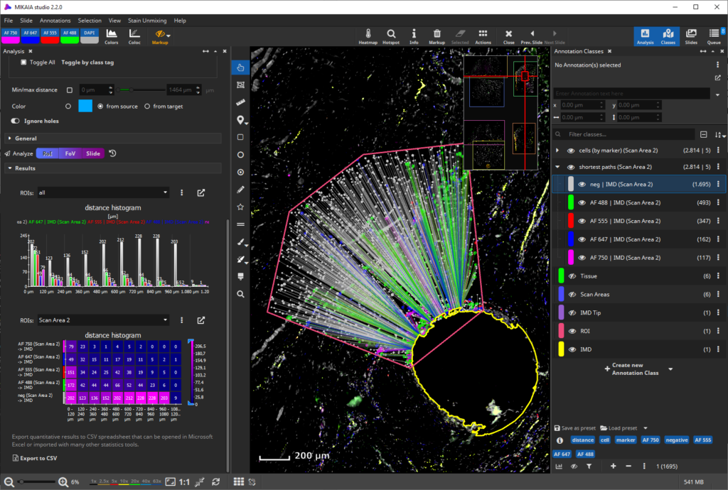
Use Case
Proximity Analysis: Quantifying Cell Subpopulations in Vicinity of Intratumoral Microdevice (IMD)
Use case from MIKAIA® users at Brigham and Women’s hospital, Harvard Medical School
– new: Feb 12th 2025 –
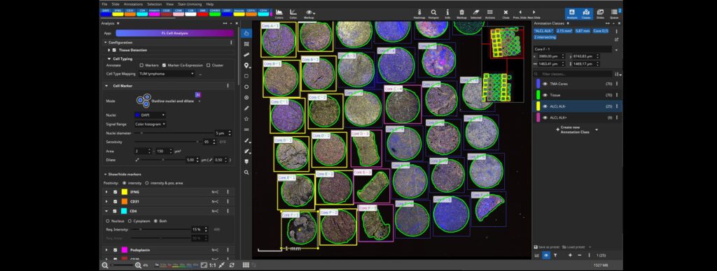
Use Case
Programming-free Single Cell Analysis of CODEX TMA with MIKAIA®
Poster at Europ. Spatial Bio conference in Dec 2024, Berlin, together with TU Munich
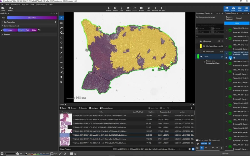

Workflow
MIKAIA Plugin API: “Plug in your own AI”
How bioinformaticians plug in their own Python (or other) scripts into the MIKAIA® app center
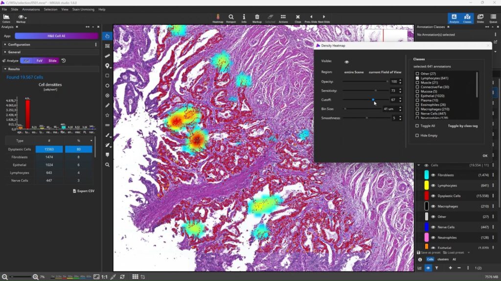
Use Case
Build H&E Analysis using multiple AIs
Video 1: Quantify cells in colon mucosa
Video 2: Detect tumor infiltratring lymphocytes (TILs)
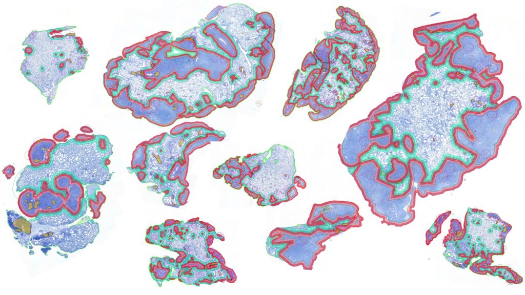
Use Case
Differential IHC Cell Detection by ROI
Video: Count IHC+ cells and compare inside vs outside metastasis
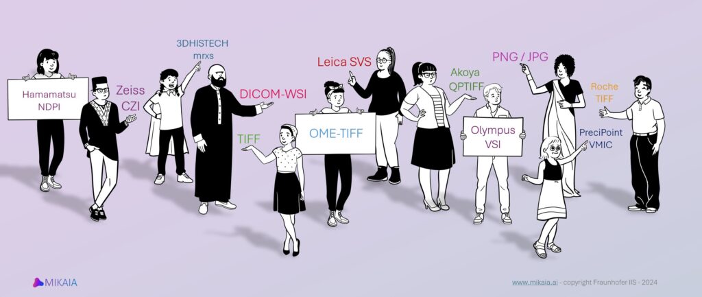
Concept
WSI and Annotation File Format I/O Support in MIKAIA
MIKAIA® supports various I/O formats, find out which ones
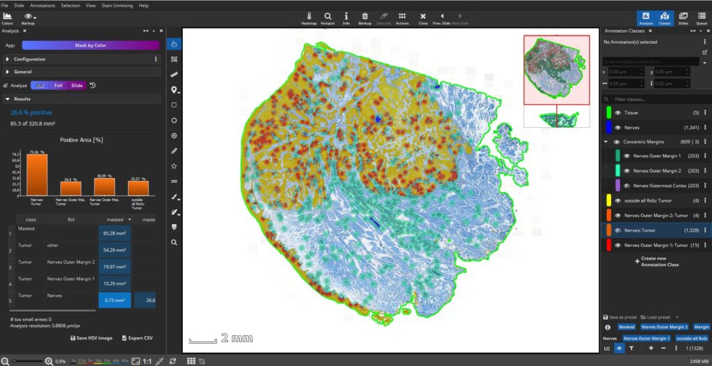
Use Case
Quantifying Perineural Invasion in Duplex IHC
… using the Mask-by-Color App and stain deconvolution
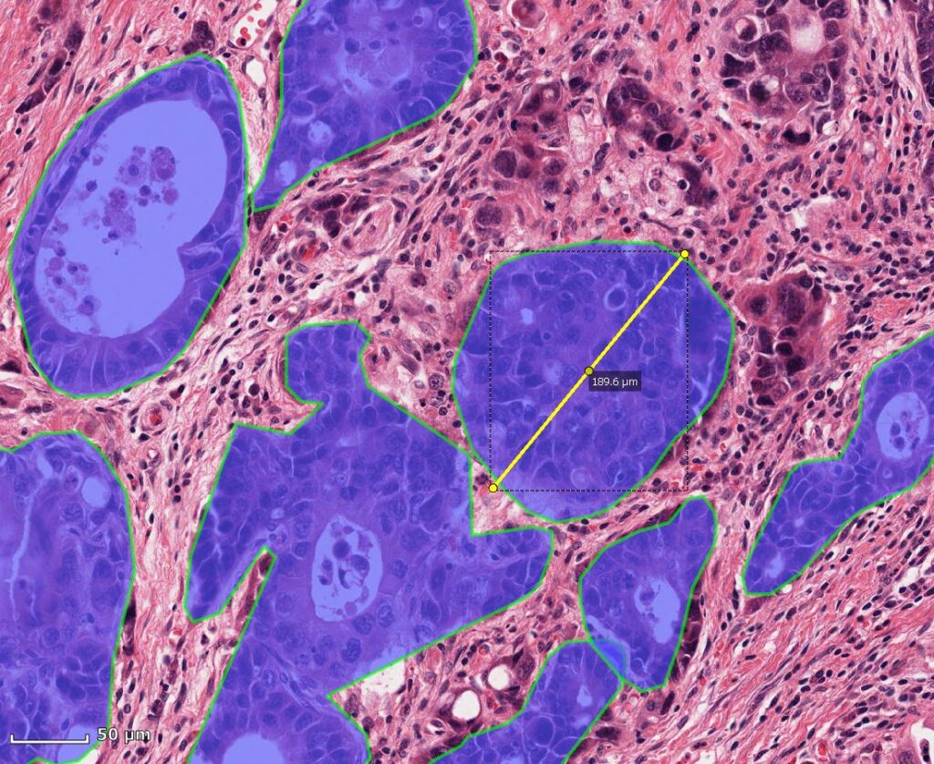
Concept
Annotation Concepts in MIKAIA for Whole-Slide-Images
Learn about MIKAIA® annotation concepts, tools, etc.
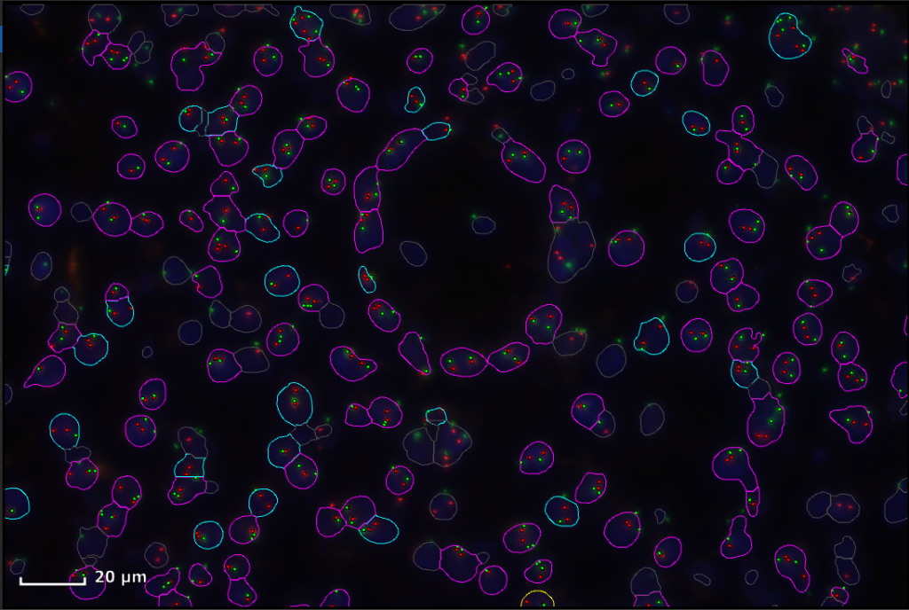
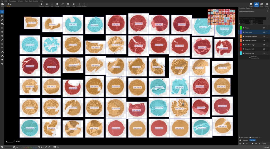
Use Case
IHC Profiler – Assessing new IHC assay using multi-organ TMA
Computing intensity and positive area per core
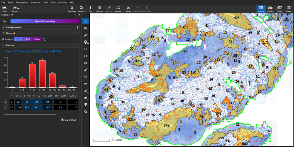
Workflow
Spatial Clustering App
Grouping spatially adjacent cells into clusters and computing histogram of cluster sizes
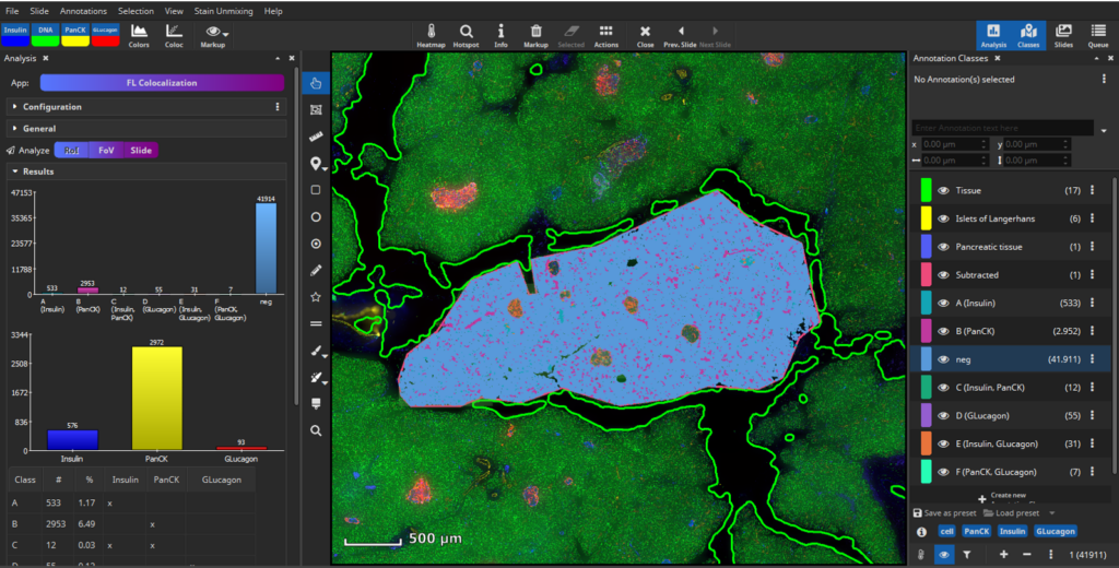
Use Case
MIKAIA-Analysis of Human Pancreas 4plex Scanned with NanoString GeoMx DSP
Cell segmentation & cell-cell-connections
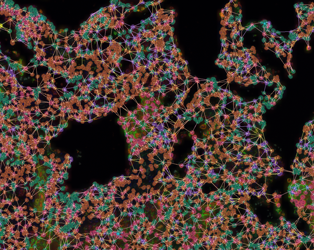
Use Case
MIKAIA-Analysis of Human Tonsil 15plex Imaged with Akoya PhenoCycler-Fusion
Cell segmentation & cell-cell-connections
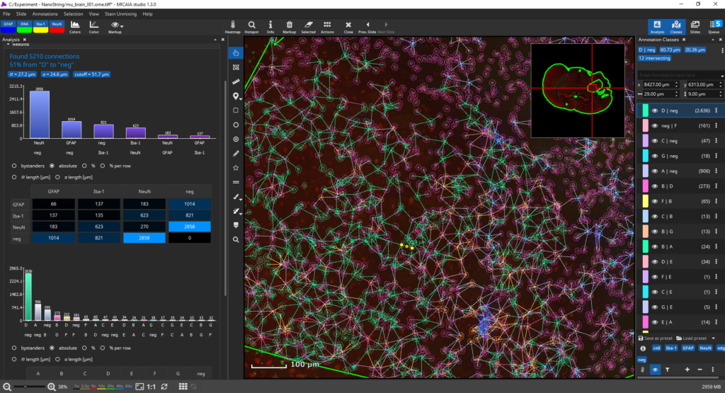
Workflow
FL Cell Analysis App
AI segmentation of nuclei or membrane. Phenotype by marker, by co-expression, or by clustering
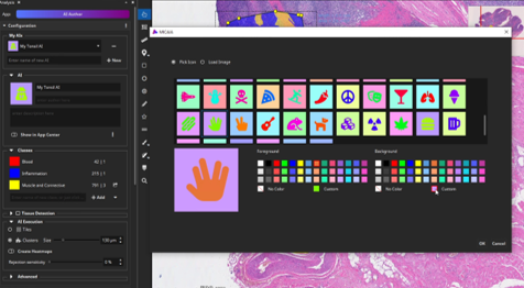
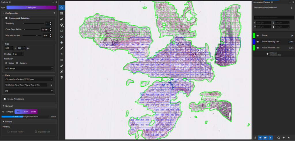
Workflow
Tile Export App
Exporting tiles, optionally with masks, to train AIs outside of MIKAIA®
available in MIKAIA® lite

Workflow
IHC Cell Detection App
Quantifying IHC+ and IHC- cells in most nuclear, membranous, or cytoplasmic stains. Finding hotspots, cluster, group by ROI.


